e-Anatomy: Your Lifeline Human Anatomy Atlas for Medical Mastery
Staring at the cadaver in my third-year dissection lab, cold sweat traced my spine as I struggled to identify the brachial plexus branches. That haunting uncertainty vanished when I discovered IMAIOS e-Anatomy during residency. This isn't just another app—it's the digital mentor I wish I'd had when textbooks left gaps in my understanding. Designed for physicians, radiologists, and students drowning in anatomical complexity, it transforms overwhelming detail into navigable knowledge. The moment I zoomed into my first cross-sectional MRI slice with labeled vasculature, I felt the visceral relief of finally grasping spatial relationships that flat diagrams never conveyed.
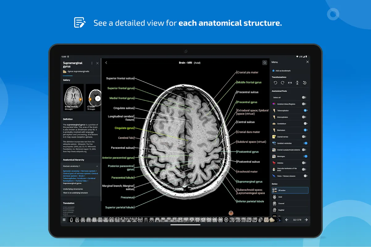
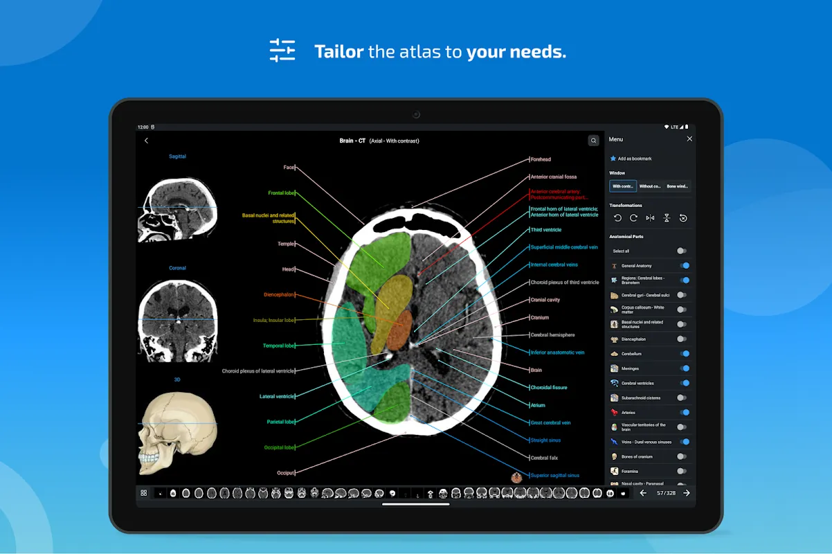
Interactive Image Exploration became my daily ritual. During tumor board meetings, dragging through axial/coronal/sagittal slices felt like physically peeling tissue layers. When consulting on a tricky femoral neck fracture, pinching to zoom revealed trabecular patterns I'd previously missed—my attending's approving nod when I pointed out the fracture line was pure validation.
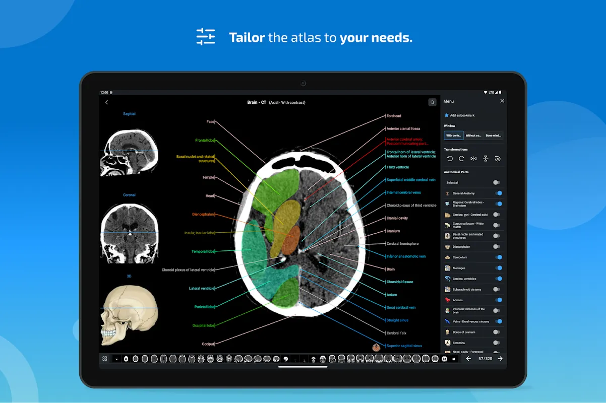
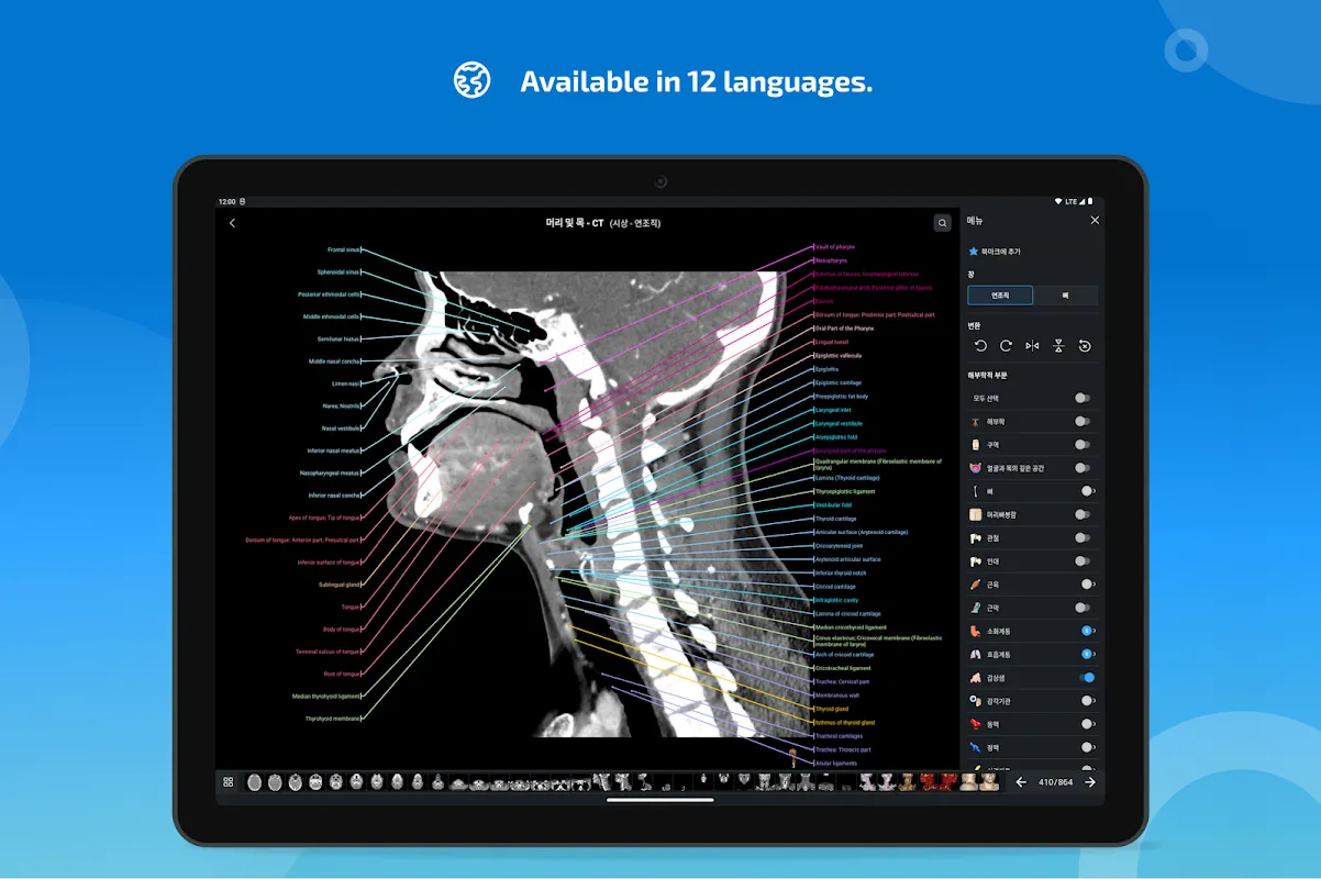
Intelligent Labeling System saved me during night shifts. One rainy ICU night, a septic patient needed emergency central line placement. Tapping the supraclavicular fossa label instantly illuminated the subclavian vein pathway on my tablet. The tactile confirmation of anatomical landmarks under my gloved fingers synced perfectly with the app's annotations—turning panic into precision.
Polyglot Terminology Database bridged language barriers in our multicultural hospital. When a Spanish-speaking patient described referred pain, switching to Latin Terminologia Anatomica helped our team align faster than any translator. Seeing "nervus ischiadicus" highlighted while palpating sciatic nerve pathways created cognitive anchors no textbook could match.
Offline Index Navigation proved invaluable during transatlantic flights. Cramming for my radiology boards at 30,000 feet, searching "circle of Willis" instantly pulled up angiograms and 3D reconstructions. Tracing arterial segments on the dimmed screen while turbulence rattled the cabin, I realized this was anatomy education unchained from Wi-Fi dependencies.
Tuesday 3 AM in the reading room: Coffee gone cold beside my PACS station. A resident paged me about ambiguous adrenal gland margins on a trauma scan. Pulling up e-Anatomy's dissection series, we toggled between sagittal views and actual cadaver photos. The way the app's coronal slices mirrored our CT images brought collective "aha" murmurs from the sleep-deprived team.
Saturday morning sunlight floods my home office as I prep Monday's lecture. Rotating a 3D skull model on my tablet while sketching zygomatic arch pathways on paper, the app's multilingual labels help me annotate slides for international students. The seamless transition from screen to whiteboard makes complex craniofacial anatomy feel unexpectedly intuitive.
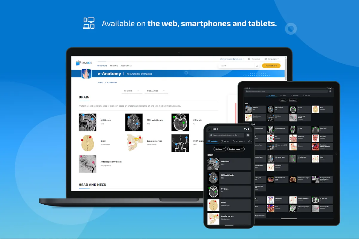
The brilliance? Launching faster than opening a physical atlas—critical when seconds count during procedures. Having 26,000+ images in my pocket outweighs the $124.99 annual fee, though I wish restored purchases didn't require periodic online checks. While the sheer volume of labels occasionally overwhelms new users, nothing compares to tapping a structure during surgery and seeing it glow on screen. For anyone navigating the human body's labyrinth—from first-year students dissecting cadavers to surgeons planning reconstructions—this is the uncompromising reference that grows more indispensable with every use.
Keywords: anatomy atlas, medical imaging, radiology reference, clinical education, anatomical labeling