MRI Essentials: Orthopedic MRI Mastery in Your Pocket
Staring at that ambiguous knee MRI last Tuesday, sweat beading on my forehead as the clock ticked toward clinic hours, I finally understood why seasoned radiologists kept whispering about this app. MRI Essentials didn't just rescue me from diagnostic uncertainty - it fundamentally changed how I approach musculoskeletal imaging. Created from Dr. Fischer's renowned MR-NOTES legacy and refined through a decade of clinical validation, this isn't another bulky digital textbook. It's the swift, precise mentor I wish I'd had during residency, now condensed into a device that fits in my lab coat pocket.
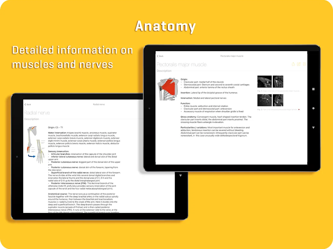
Concise Visual Reference transforms overwhelming protocols into actionable knowledge. That moment I first swiped through the basic version's illustrated modules - simple ink drawings beside bullet-point explanations - felt like removing earplugs I didn't know I wore. During hectic morning rounds last month, the carpal tunnel syndrome diagrams allowed me to explain scapholunate ligament tears to interns in ninety seconds flat, their nodding faces confirming the clarity even junior clinicians appreciate.
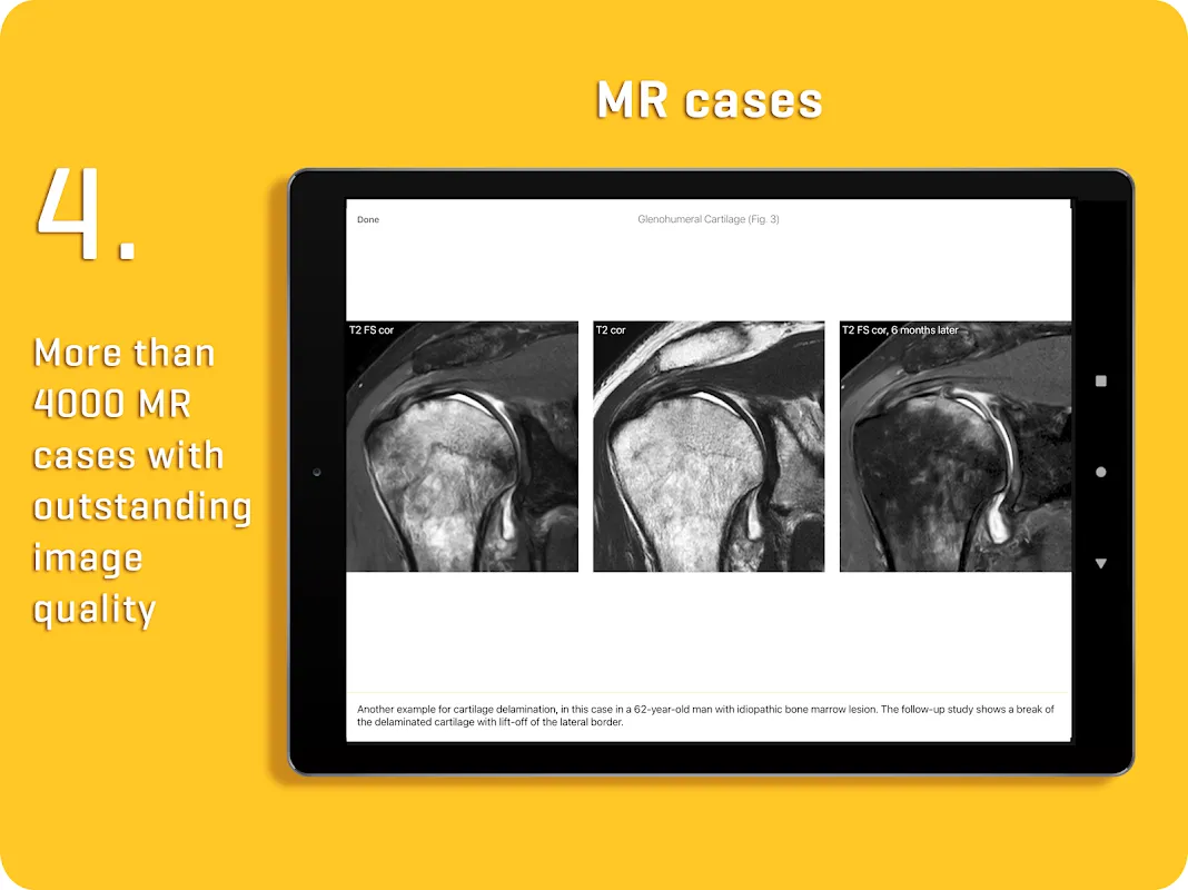
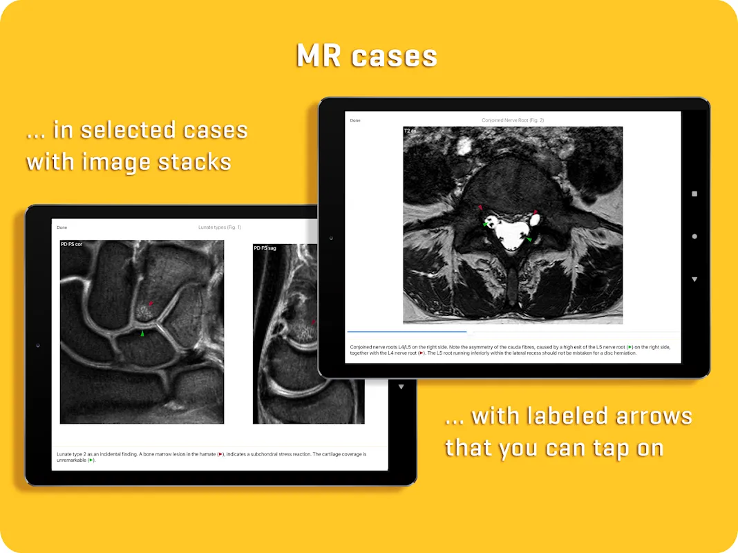
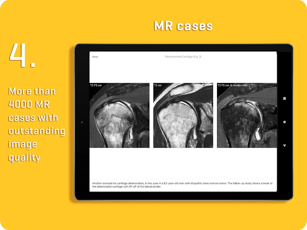
Pro's Living Atlas with over 4,000 cases elevates theoretical knowledge into clinical instinct. I recall the rainy Thursday when a teenager's atypical hip pain stumped our entire team. Zooming into the app's pristine coronal STIR sequences revealed subtle femoral head edema I'd have missed otherwise. The tactile sensation of fingertips spreading across the tablet screen to enlarge a tibial stress fracture case last week created muscle-memory learning no textbook could replicate. Each case feels like walking alongside Dr. Fischer during his teaching rounds.
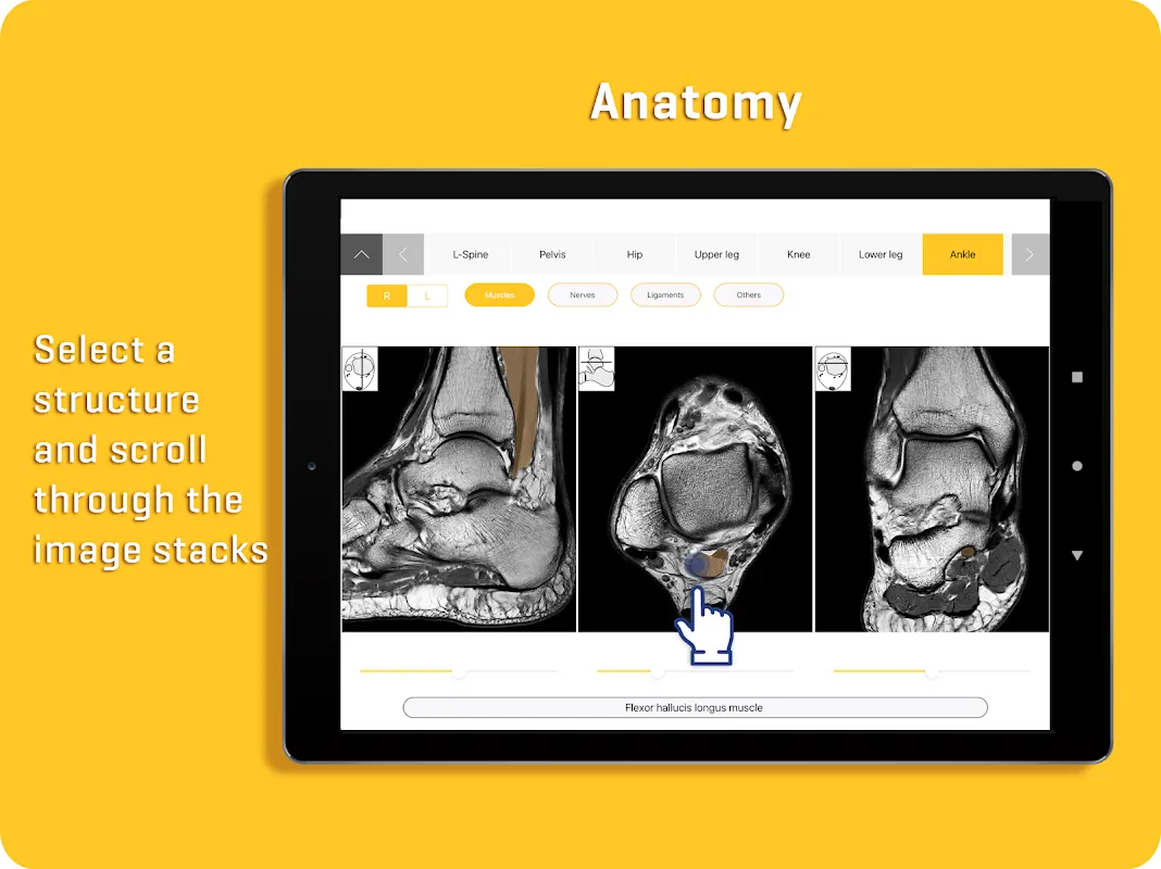
Dual-Path Navigation anticipates our clinical workflow dilemmas. Pre-op consultations now involve tapping between concise teaching notes during patient questions, then seamlessly switching to the case gallery when discussing surgical visuals. During yesterday's MRI protocol meeting, flipping from basic meniscal tear schematics to advanced post-op comparison scans silenced the usual projector-fumbling chaos. The interface remembers my last position like a considerate colleague saving your page in a shared journal.
Tuesday 7:03 AM, reading room coffee steaming beside elbow. My index finger traces the subtle cortical irregularity on a humeral scan while the app's corresponding case glows on the secondary monitor. The pre-dawn quiet amplifies each swipe through proximal biceps tendon examples - no keyboard clicks, no paper rustle - just the silent certainty of matching pathology patterns. Later that afternoon, sunlight stripes across the conference table as residents lean in, their tablets mirroring my screen showing Bankart lesion variants. That shared "aha" when the app's surgical correlation images appear still gives me teaching chills.
The brilliance? Launching faster than PACS during emergencies. That terrifying on-call moment when the ER phoned about possible spinal compression? MRI Essentials loaded before I finished saying "sagittal T2." But I'd sacrifice coffee for a week if they'd add cross-referenced biomechanics notes - like last month when that ballet dancer's complex ankle instability had me juggling three apps. Still, watching moonlight reflect off my iPad during midnight studies, I realize no resource balances depth with accessibility better. For orthopedists drowning in imaging data yet starved for time, this isn't just helpful - it's practice-saving.
Keywords: orthopedic MRI, radiology reference, medical imaging, diagnostic tool, clinical teaching
