Circulatory System 3D Anatomy: Master Blood Pathways with Interactive Heart Visualization
Staring at flat textbook diagrams during my physiology rotation, I felt like an explorer lost without a compass. That sinking frustration vanished when I discovered this app. Suddenly, the intricate highways of our blood vessels became tangible landscapes I could navigate with my fingertips. Designed for medical learners yet accessible enough for curious minds, this tool transformed my understanding of vascular pathways from abstract concepts to living structures.
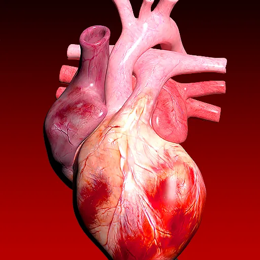
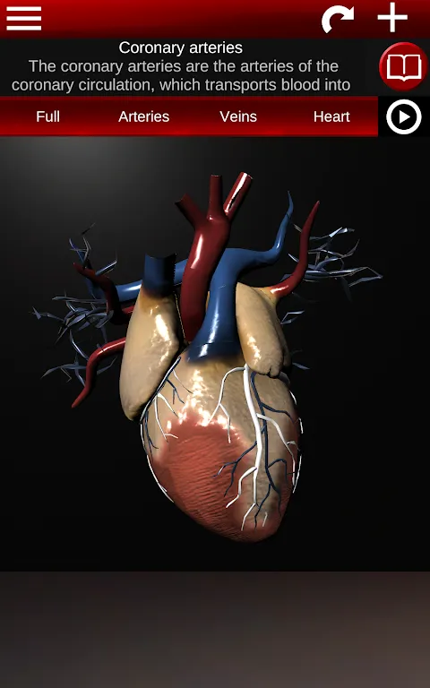
The moment I zoomed into capillary networks felt like peering through a microscopic lens. During hematology studies, pinching to magnify those delicate vessels revealed branching patterns no illustration could capture. My fingers instinctively traced oxygen exchange points, the tactile feedback creating muscle memory that later surfaced during practical exams. Rotating the 3D heart model became my nightly ritual. After clinical rounds, I'd spin the pulsating organ in midnight darkness, observing valve movements from oblique angles. The realism struck me when comparing it to intraoperative videos - identical mitral valve mechanics fluttering beneath my touch.
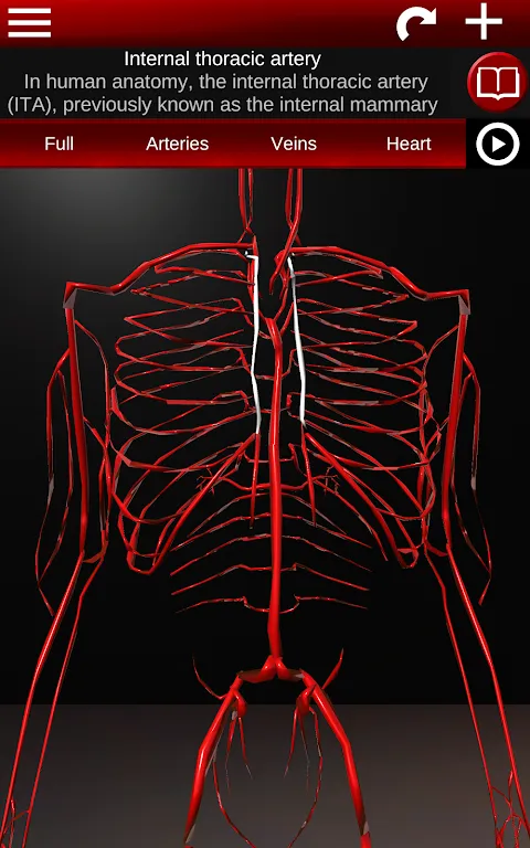
What truly reshaped my study approach was the toggle labeling feature. Preparing for anatomy viva, I'd hide all terms and quiz myself on cadaveric specimens. The panic of blanking on basilic vein pathways dissolved when tapping the screen revealed answers, the relief physical as shoulders unlocked. That selective visibility proved equally powerful during patient consultations. Last Tuesday, explaining coronary circulation to a nervous gentleman, I hid complex terminology, focusing purely on blood flow visualization. His tense expression softened as he pointed at the rotating arteries, finally grasping his blockage's location.
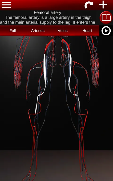
Language flexibility addressed struggles I hadn't anticipated. When assisting French exchange students, switching terminology mid-demonstration bridged our knowledge gaps instantly. Their spontaneous "Ah, l'artère fémorale!" when recognizing the femoral artery confirmed how seamlessly multilingual support aids collaborative learning. Beyond academics, I've used the beating heart animation during CPR workshops. Freezing the cycle to show ventricular filling stages made compression timing click for trainees in ways manikins never could.
Pre-dawn study sessions reveal the app's strengths and limits. The instant launch saves precious minutes during hospital downtime - quicker than scrubbing in for emergency cases. Yet reviewing posterior tibial veins during a thunderstorm, I wished for adjustable lighting to counteract screen glare. While artery labeling is impeccable, some deep calf veins lack depth cues that would help surgical planning. Still, watching interns cluster around my tablet during lunch breaks proves its unmatched value.
For nocturnal learners craving spatial understanding, this is essential. Whether you're memorizing aortic branches at 3 AM or explaining angina to grandparents, it turns vascular mysteries into intuitive journeys. Just keep a stylus handy - you'll be sketching pathways in your sleep.
Keywords: anatomy, circulatory system, medical education, 3D visualization, heart model