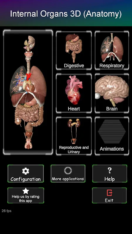
Internal Organs in 3D Anatomy: Interactive Learning for Medical Students and Curious Minds
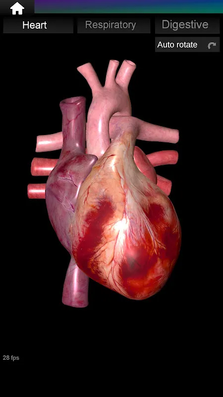
Staring at flat textbook diagrams left me frustrated during my physiology certification prep. Flat images couldn't convey how the trachea branched into bronchi or how the intestines coiled. That changed when I discovered this app during a late-night study crisis. Within minutes of exploring its rotating heart model, spatial relationships clicked. Now I recommend it to every healthcare trainee needing visceral understanding beyond memorization.
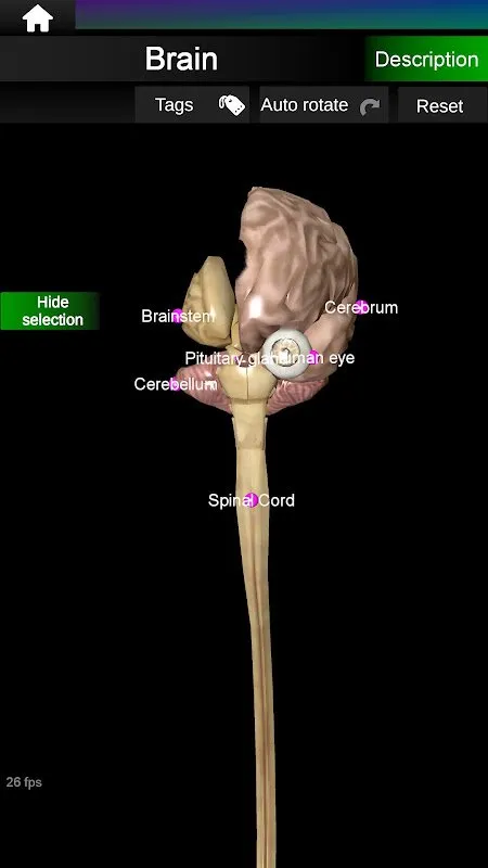
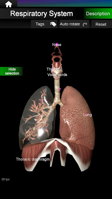
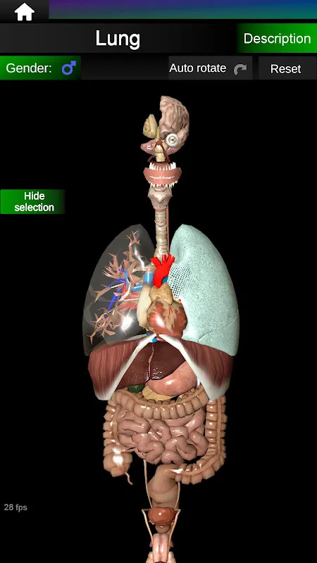
True 3D Exploration became my game-changer. Pinching to zoom into the cerebellum's fissures while studying for neurology finals, I noticed details invisible in lab specimens. Rotating the lungs during a respiratory module revealed hidden posterior lobes. That tactile control created muscle memory far faster than static images ever did.
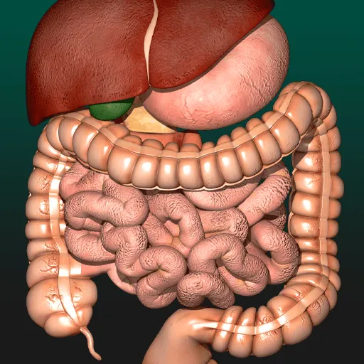
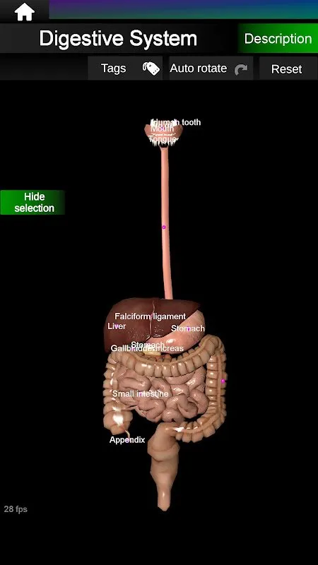
System-Specific Animations transformed abstract concepts. Watching peristalsis ripple through the digestive tract while prepping for a tutoring session made me gasp. Suddenly, textbook descriptions of muscular contractions became living processes. When explaining aortic valve function to my niece, the beating heart visualization held her attention better than cartoons.
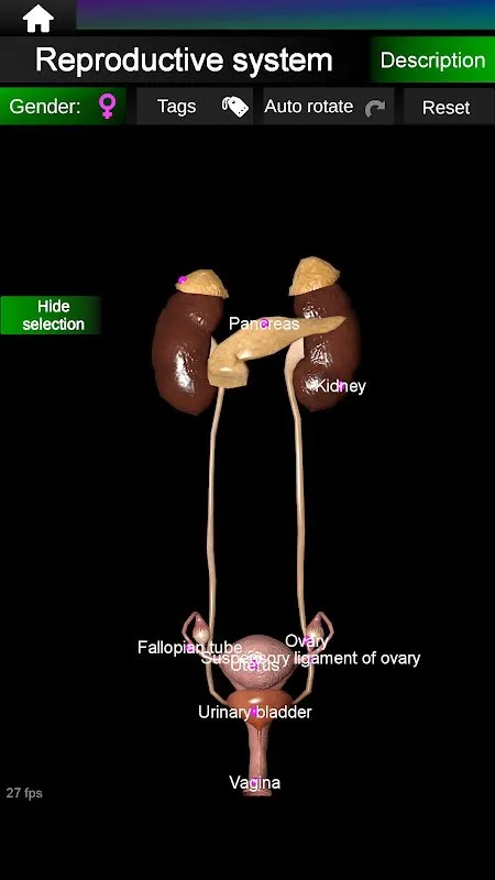
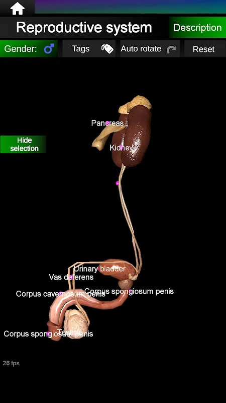
Dynamic Comparison Tools saved me during obstetrics rotations. Switching between male and female reproductive models helped clarify uterine positioning for a patient consultation. Hiding layers to isolate the brainstem then gradually revealing surrounding structures built my confidence before a neurosurgery observation.
Multilingual Accessibility proved unexpectedly vital. During an international medical conference, a Russian researcher struggled with English organ descriptions. Switching the interface to his native language sparked an animated discussion about hepatic structures over coffee. That seamless transition fostered collaboration no textbook could match.
Tuesday 3 AM: Deadline panic set in before my digestive system presentation. Moonlight illuminated the phone screen as I rotated a 3D stomach model. Tracing gastric folds with my fingertip, the texture mapping felt eerily real. Suddenly, the animation showed chyme entering the duodenum – that visceral understanding sparked my final slide design.
Saturday clinic waiting room: A teenager nervously asked about his asthma. Pulling up the respiratory system, we zoomed into bronchioles together. His fingers trembled slightly rotating the model, but seeing inflamed airway simulations sparked genuine questions. That shared exploration built trust faster than medical jargon.
Post-surgery recovery: My grandfather struggled to comprehend his bypass procedure. With device narration in Spanish, we explored coronary arteries layer by layer. Watching his calloused finger trace the aorta's curve, I saw fear transform into engagement. That moment justified every megabyte of storage space.
The pros? Launch speed rivals emergency pager apps – crucial during impromptu patient explanations. Model accuracy consistently impresses professors; I've spotted details matching cadavers. But heavy animations drain batteries during marathon study sessions. Occasional texture glitches in intestinal villi require restarting. Still, these pale against its brilliance. Essential for visual learners drowning in textbooks, yet intuitive enough for curious highschoolers. Keep it installed beside your medical references.
Keywords: anatomy, 3D models, medical education, interactive learning, human body