Rotating Kidneys Saved My Exam
Rotating Kidneys Saved My Exam
My palms were sweating onto the library desk as I squinted at yet another 2D diagram of nephrons. That cursed renal pyramid looked like a flat triangle - where were the tubules wrapping around it? How did the blood vessels penetrate the cortex? I'd failed two quizzes already, and Professor Davies' warning echoed: "If you can't visualize it, you can't diagnose it." Desperation tasted like stale coffee when I slammed the textbook shut at 3 AM.
The digital cadaver
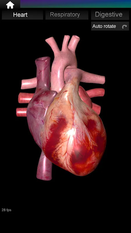
That's when Marco from surgery rotation texted: "Stop torturing yourself. Try the 3D anatomy thing." Skepticism curdled in my throat as I downloaded it - another gimmicky app? But loading the kidney model made me gasp. Suddenly I was holding a pulsating organ with volumetric lighting revealing every minor calyx. My finger spun it like a cosmic marble, zooming until Bowman's capsule enveloped my screen. For the first time, I saw how the afferent arteriole snaked into the glomerulus like a red vine. The app used photogrammetry from real cadavers - no wonder the tissue texture looked disturbingly authentic when I virtually dissected the renal pelvis.
Rotating the model with trembling fingers, I finally understood why our patient's ureter obstruction caused hydronephrosis. The pelvis wasn't some abstract sac - it was a delicate funnel crumpling under pressure. When I tapped the hypertension layer, arteries constricted before my eyes, showing ischemic areas in sickly blue. This wasn't studying; it was revelation. I spent hours exploring angles textbooks never showed - peering upward through the urethra, watching urine flow in ghostly animation. The haptic feedback vibrated when I "nicked" an artery during dissection practice. My trashcan filled with coffee cups as dawn bled through the blinds.
When pixels failBut fury struck during the adrenal gland module. The model glitched, superimposing cortex over medulla until it looked like a cubist nightmare. I nearly threw my tablet when the app crashed after 45 unsaved minutes of tracing arterial supply. And why did the pancreatic model feel like plasticine compared to the hyper-real kidneys? Developers clearly played favorites with organs. Still, I returned like a masochist because even flawed 3D beat perfect 2D. That night I dreamt in cross-sections.
Exam day arrived with monsoon rains. When question 17 demanded identifying renal structures in a CT scan, panic seized me - until I mentally rotated my digital kidney. I traced the path from major calyx to urethra like following subway lines. The invigilator frowned at my grinning. Results came Friday: 94% in renal module. Professor Davies scrawled "Finally visualized!" in red ink. Now I smirk when classmates stress over flat diagrams. Sure, the app crashes if you breathe wrong, and its subscription fee should fund a kidney transplant. But when you're wrist-deep in virtual fascia, suddenly memorizing becomes knowing.
Keywords:Internal Organs in 3D Anatomy,news,medical visualization,interactive learning,anatomy education


